Abdominal Anatomy : The abdominal anatomy stock illustration. Illustration of ... - Introduction to sonographic abdominal anatomy.
7:30 PM
Abdominal Anatomy : The abdominal anatomy stock illustration. Illustration of ... - Introduction to sonographic abdominal anatomy.. Become familiar with the anatomical divisions by exploring the world's most advanced 3d anatomy platform in complete anatomy. Divided into 9 regions by two vertical and two horizontal imaginary planes. Anatomy of the abdominal wall, inguinal region & hernias, hernias, gut and peritoneal cavity. Sciency root words make anatomical parts harder to memorize. The abdomen (colloquially called the belly, tummy, midriff or stomach) is the part of the body between the thorax (chest) and pelvis, in humans and in other vertebrates.
Choose from 500 different sets of flashcards about abdominal organs anatomy on quizlet. • in this module, we will explore basic abdominal anatomy identifiable with common imaging modalities. We'll identify as many organs as we can, see how they fit into. A collection of articles covering abdominal anatomy, including abdominal wall anatomy and abdominal cavity anatomy. Abdominal cavity, largest hollow space of the body.
Abdominal Anatomy Surface - Surface Anatomy of the ... from medicinebtg.com • in this module, we will explore basic abdominal anatomy identifiable with common imaging modalities. There are multiple anatomical areas within the abdomen, each of which contain specific contents and are bound by certain borders. A good amount of area is covered by the abdominal wall. • the abdomen consists of: The above lines intersect and divide the abdomen into nine regions (clockwise from the top) We will wrap up with an overview of several abdominal diseases that might all present themselves with pain. We'll identify as many organs as we can, see how they fit into. The anterior abdominal wall (figs.
Most students entering ultrasound have some basic understanding of anatomy. 6 write the origin, insertion and nerve supply of muscles of anterior abdominal wall. Lee moffitt cancer center & research institute in. This muscle forms the anterior and lateral abdominal wall. We're going to take apart a plastic anatomy model and see what we can find in the abdomen. The epigastric vessels (long arrow); Identify abdominal anatomical structures in a variety of medical imaging platforms. Two layers in abdomenfatty superficial layer (camper's fascia)deeper membranous layer (scarper's fascia). The anterolateral abdominal wall formed of 4 layer skin, fascia, muscles, and peritoneum. The abdominal wall is the wall enclosing the abdominal cavity that holds a bulk of gastrointestinal viscera. Sectional anatomy the sonographer must have a working knowledge of anatomical structures with particular attention to spatial relationships within the body. Identify some abdominal pathology on medical images. • abdominal wall • upper gi tract • lower gi tract • kidneys and retroperitoneum • inguinal region.
Abdominal anatomy seen on ct. The quadratus lumborum muscle (black arrow). Laterally by the midaxillary line; A collection of articles covering abdominal anatomy, including abdominal wall anatomy and abdominal cavity anatomy. The transversus abdominis muscle is the deepest of the abdominal muscles, lying internally to the internal abdominal obliques.
Abdominal muscles | Golf Loopy - Play Your Golf Like a ... from cloud2.golfloopy.com Introduction to sonographic abdominal anatomy. Identify abdominal anatomical structures in a variety of medical imaging platforms. The above lines intersect and divide the abdomen into nine regions (clockwise from the top) Abdominal cavity, largest hollow space of the body. Most students entering ultrasound have some basic understanding of anatomy. We'll identify as many organs as we can, see how they fit into. The linea alba (open arrowhead); The xiphoid process and costal.
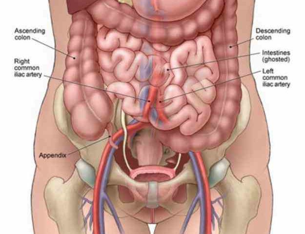
Anatomical structures of the abdomen and pelvis are visible as interactive labeled images.
The abdominal divisions should be used in conjunction with other diagnostic approaches in order to accurately diagnose a patient's condition. Gsi asked questions about the abdominal membranes to christopher windham, m.d. Demonstrate comprehension of core abdominal anatomy. Anatomical structures of the abdomen and pelvis are visible as interactive labeled images. Sectional anatomy the sonographer must have a working knowledge of anatomical structures with particular attention to spatial relationships within the body. Most students entering ultrasound have some basic understanding of anatomy. Identify abdominal anatomical structures in a variety of medical imaging platforms. But with the use of smart technology, you can learn faster and master abdomen anatomy in no time! Divided into 9 regions by two vertical and two horizontal imaginary planes. Identify some abdominal pathology on medical images. A good amount of area is covered by the abdominal wall. We'll identify as many organs as we can, see how they fit into. Sciency root words make anatomical parts harder to memorize.
Introduction to sonographic abdominal anatomy. Abdominal anatomy seen on ct. Anatomical structures of the abdomen and pelvis are visible as interactive labeled images. Its upper boundary is the diaphragm, a sheet of muscle and connective tissue that separates it the abdominal organs are supported and protected by the bones of the pelvis and ribcage and are covered by the greater omentum, a fold of peritoneum. Two layers in abdomenfatty superficial layer (camper's fascia)deeper membranous layer (scarper's fascia).
The abdominal anatomy stock illustration. Illustration of ... from thumbs.dreamstime.com Divided into 9 regions by two vertical and two horizontal imaginary planes. Learn about abdominal organs anatomy with free interactive flashcards. The above lines intersect and divide the abdomen into nine regions (clockwise from the top) Radiology basics of abdominal ct anatomy with annotated coronal images and scrollable axial images to help medical students and junior doctors learning anatomy. The abdominal region is supported by the anterior and posterior abdominal wall that supports the viscera and maintains the posture where there's no bony support. Compare and contrast the different medical imaging modalities presented in the tutorials. You will learn the anatomical basis of pain and how to apply this knowledge in the diagnostic process. 6 write the origin, insertion and nerve supply of muscles of anterior abdominal wall.
Become familiar with the anatomical divisions by exploring the world's most advanced 3d anatomy platform in complete anatomy.
Radiology basics of abdominal ct anatomy with annotated coronal images and scrollable axial images to help medical students and junior doctors learning anatomy. The quadratus lumborum muscle (black arrow). The abdominal divisions should be used in conjunction with other diagnostic approaches in order to accurately diagnose a patient's condition. 6 write the origin, insertion and nerve supply of muscles of anterior abdominal wall. Sectional anatomy the sonographer must have a working knowledge of anatomical structures with particular attention to spatial relationships within the body. Learn about abdominal organs anatomy with free interactive flashcards. • in this module, we will explore basic abdominal anatomy identifiable with common imaging modalities. Divided into 9 regions by two vertical and two horizontal imaginary planes. The transversus abdominis muscle is the deepest of the abdominal muscles, lying internally to the internal abdominal obliques. • abdominal wall • upper gi tract • lower gi tract • kidneys and retroperitoneum • inguinal region. Identify some abdominal pathology on medical images. Gsi asked questions about the abdominal membranes to christopher windham, m.d. Therefore, a firm grasp of abdominal anatomy is necessary to effectively diagnose and treat patients.